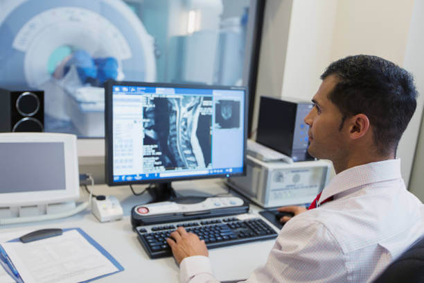
Do you need an MRI arthrogram? Read on to learn more about what to expect during this procedure.
Why You Might Need an Arthrogram
Arthrographic images help doctors evaluate the structure of a joint along with its function to determine if there is a need for treatment. Your doctor may prescribe an arthrogram as part of their diagnosis of the soft tissue structures inside your shoulder, hip, knee or another joint. Some of the main reasons to do this test include:
- Persistent and unexplained joint discomfort or chronic pain
- Loss of motion
- Changes in the way the joint works
- Persistent and unexplained swelling
- Problems in the ligaments, cartilage, tendons and other soft tissues of the joint
- Damage from repeated dislocations
- To check prosthetic joints
- To search for loose bodies within the joint
There may be other reasons your doctor recommends arthrography. The imaging procedure, along with the contrast dye, provides a clear image of the soft tissue in the joint. With these pictures, your doctor can clearly see if there is an underlying issue, condition or cause for your painful symptoms. This information helps your doctor determine a course of treatment or even recommend a surgical procedure such as joint replacement.
Common Uses of Arthrograms
Knee and other joint arthrograms are used to help your doctor evaluate the structures and function of a particular joint. If they view any alterations to the joint, this procedure helps them determine the best form of treatment for your condition.
Some of the most common uses for arthrograms are to identify abnormalities within your joints, for example:
- Shoulder: Determine where the joint is unstable or find a suspected tendon tear.
- Hip: Show if there is a tear in the rim of the joint — the cartilage labrum.
- Wrist: Discover any tear of the small ligaments.
- Knee: View any issues in the meniscus or discover cartilage knee lesions.
- Ankle: Study and evaluate ankle sprains and other issues.
- Elbow: Investigate pain in patients who repeatedly use their elbow, such as throwing athletes.
There are many other situations where your doctor may feel that an arthrogram could provide the additional information they need to help determine the best course of treatment.
In some cases, arthrogram procedures have a therapeutic use. Arthrograms can be used to correctly position a needle within a joint so that medication or an anesthetic can be injected to decrease joint-related pain or inflammation.
Benefits of Arthrograms
There are many unique benefits that arthrographic images provide to doctors and medical specialists, including:
- Arthrograms are a minimally invasive imaging technique that presents little risk to the patient.
- The use of contrast material improves the quality of the MRI or CT to more accurately show damage in and around the internal structure of your joint.
- They do not expose patients to ionizing radiation.
- Arthrograms are particularly effective for detecting disease, tears, lesions and other damage to the structures within the joints such as the ligaments, tendons, cartilage and labrum.
- Arthrography allows for the discovery of abnormalities, such as those obscured by bone, that may not be visible with other imaging methods.
How to Prepare for an Arthrogram
Before the arthrogram procedure, our team at Health Images will ensure you’re fully aware of what to expect and any potential risks involved. You’ll also be able to ask any questions you may have. You will need to sign a consent form permitting us to perform this procedure, and we’ll need to know ahead of time if you have allergies to latex, tape, anesthetics, contrast dye, iodine or any medicines. You should also let us know about any medication or supplements you’re taking, or if you believe you may be pregnant.
You don’t need to worry about a special diet or restricting activity before the procedure. You may also receive other instructions on the day of your arthrogram procedure.
What Happens During and After an Arthrogram?
When you arrive at Health Images, you may need to wear medical scrubs (top and pants) or a hospital gown during the procedure or remove all clothing around the joint being tested. You’ll also need to remove all metal objects, including jewelry.
The process of taking arthrogram images may vary, but in general, here is what you can expect during the procedure:
- You will lay on a table in the examination room.
- Before the contrast material is injected, X-rays may be taken to later compare with the arthrogram’s results.
- The area around your joint will be covered, and the skin above the joint cleaned and numbed.
- Sometimes, fluid is removed from around the joint using a longer needle.
- CT, Fluoroscopy or ultrasound are used to guide a needle into the joint to inject the contrast dye.
- To spread the dye, you may be asked to move your joint.
- Using x-ray, MRI, CT scans or fluoroscopy, a machine will take images of the joint in different positions.
The arthrogram procedure can last anywhere from 30 minutes to two hours, depending on the type of imaging being used.
Once the procedure is complete, here’s what to expect afterward:
- You will receive specific instructions on the movement of your joint, activity restrictions, pain medication that’s safe and adverse symptoms to watch out for, like fever, pain, swelling or drainage around your injection site.
- It’s typical to rest your joint for several hours once the arthrogram is finished.
- Mild swelling is normal and can be treated with a cold compress. However, if it continues, notify your physician.
- Knee joints may be wrapped for several days after a knee arthrogram.
- You may notice clicking or cracking sounds when you move your joint for a few days. Don’t be alarmed, as this is normal and should resolve after a few days.
Visit a Health Images Location
If you’re in need of an MRI arthrogram or another diagnostic imaging procedure, turn to the trusted team at Health Images. With imaging centers throughout the Denver area and beyond, you’re sure to find a location near you.
Find Your Nearest Location


