How are X-rays Produced?
You might be familiar with x-rays or may have had one at some point, but what exactly does the process entail? A lot goes into an x-ray to ensure a clear, high-quality image of the area being examined, allowing the doctor to detect a potential issue and determine the right course of action.
This article aims to answer some common questions about x-rays to give you a better idea of how they work. Learn about their different uses, how they’re produced, whether they’re safe and how they use electromagnetic radiation in this comprehensive guide to x-rays.
What are x-rays?
An x-ray is a quick, painless procedure that produces images of the inside of the body. It’s a highly effective way of examining the bones and can help identify various conditions.
X-rays are commonly performed by trained specialists — radiographers — in hospital x-ray departments, but other healthcare professionals like dentists use them as well.
Use of x-rays
Healthcare specialists can examine most areas of the body with x-rays, but they’re primarily used for the bones and joints. However, they’re sometimes used to detect abnormalities in soft tissue and internal organs. X-rays can be used to detect conditions such as:
- Bone breaks and fractures
- Atypical curvature of the spine, or scoliosis
- Kidney stones
- Loose teeth, dental abscesses and other tooth-related problems
- Lung-related conditions, like lung cancer and pneumonia
- Cancerous and non-cancerous bone tumors
- Breast cancer
- Swallowing difficulties, or dysphagia
- Heart disease or heart failure
X-rays can also help guide doctors and surgeons through certain procedures. For instance, a physician might use an x-ray to guide a catheter — a thin, long and flexible tube — along an artery during coronary angioplasty, which widens narrow arteries close to the heart.
How do x-rays work?
X-rays involve radiation that passes through the body, but you can’t feel it or see it with the naked eye. As x-rays pass through the body, different body parts absorb their energy at different rates. A detector on the other side of the body detects these x-rays once they’ve passed through, then turns them into an image visible on the screen.
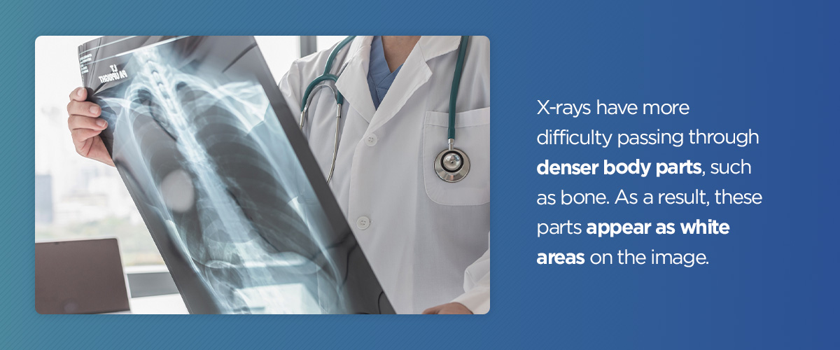
X-rays have more difficulty passing through denser body parts, such as bone. As a result, these parts appear as white areas on the image. Softer body parts like the lungs and heart are easier for x-rays to pass through, showing up as darker areas.
How are x-rays produced?
An x-ray is typically produced in an x-ray tube by accelerating electrons through a potential difference. It then directs them to a target material, such as tungsten. The radiographer can alter the voltage and current settings on the machine, manipulating the x-ray beam properties produced. Different x-ray beam spectra are used for different body parts.
The incoming electrons give off x-rays, slowing down as they pass near the nucleus. The x-ray photons created during the process can range anywhere from near zero to the energy of the electrons. An incoming electron and atom may also collide in the target, causing a vacancy in one of the atom’s electron shells. When another electron fills this vacancy, it releases a photon known as a characteristic x-ray.
With a type of x-ray machine called a computed tomography (CT) scanner, the x-ray tube generates a fan-shaped beam, which moves around the patient in a circular pattern. The CT scanner detects the x-rays electronically, using the data to reconstruct an image of the region being examined.
X-rays can also be produced with a synchrotron device. A synchrotron accelerates the electrons in a ring, steering them with magnets. Manipulating the electron beam with magnets can create intense x-ray beams. Synchrotron facilities are commonly used for research purposes.
Are x-rays safe?
It’s common to have concerns about exposure to radiation during an x-ray. Some people worry about the formation of cell mutations that may cause cancer.
In most cases, the examined area is exposed to a low level of radiation for a fraction of a second. However, the amount of radiation exposure during an x-ray depends on the organ or tissue being examined. Additionally, sensitivity to this radiation can vary by age, with children usually having higher sensitivity than adults.
Any radiation exposure can increase the risk of cancer, but the likelihood of contracting it from an x-ray is believed to be very slim. To put it into perspective, an x-ray of the chest, teeth or limbs is equivalent to a few days of natural background radiation, with less than one in a million chance of causing cancer.
Radiation exposure during x-rays is generally low, with the benefits of these tests far outweighing the risks. If you’re concerned about potential risks, don’t hesitate to talk to your radiographer or doctor beforehand.
You should also talk to your doctor if you’re pregnant or think you may be pregnant before having an x-ray. Most diagnostic x-rays pose small risks to unborn babies, but your doctor might recommend an alternative imaging test like an ultrasound.
How do x-rays use electromagnetic radiation?
Like gamma rays, radio waves, visible light and microwaves, x-rays are a form of electromagnetic radiation. X-ray photons are quite powerful, with enough energy to break up molecules. When x-rays reach a material, some pass through while others are absorbed.
In most cases, higher energy levels will result in more x-rays passing through. This penetrating power makes it possible to take internal images of the human body and other objects. With their ability to help detect and diagnose various medical conditions, including life-threatening ones, x-rays are a vital and valuable piece of technology in the medical world.
Request an x-ray appointment today
No matter why you require an x-ray, our team at Health Images is here to assist you. The world of radiology imaging is constantly evolving — that’s why we aim to maintain the most accurate and up-to-date technology, detecting and addressing potential problems as quickly as possible. You may need an x-ray for numerous reasons, including:
- Monitoring the status of an injury or disease.
- Examining a bodily area causing you pain or discomfort.
- Reviewing the progress of how a prescribed treatment or medication is working.
Whatever part of your body needs an x-ray, one of our highly trained technologists will ensure you have a pleasant and comfortable experience while getting the clearest images and necessary information to determine the next steps. Request an x-ray appointment at Health Images today!
Linked Sources:
- https://www.healthimages.com/scans-for-detecting-kidney-stones/
- https://www.healthimages.com/who-should-be-screened-for-lung-cancer/
- https://www.healthimages.com/what-are-all-the-breast-cancer-screening-options/
- https://www.arpansa.gov.au/understanding-radiation/what-is-radiation/ionising-radiation/x-ray
- https://www.healthimages.com/services/ct-scans/
- https://www.fda.gov/radiation-emitting-products/medical-imaging/pediatric-x-ray-imaging
- https://www.nhs.uk/conditions/x-ray/
- https://www.healthimages.com/services/ultrasound-sonogram/
- https://www.healthimages.com/services/x-ray/
- https://www.healthimages.com/request-an-x-ray/


