How to get the most out of your ultrasound
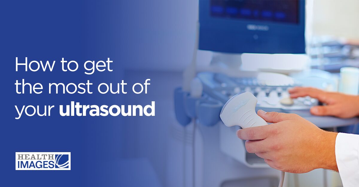 Has your physician recommended you get an ultrasound scan? If you’ve never had to get an ultrasound exam before, you may be wondering what to expect.
This guide will explain everything you need to know about ultrasound imaging, including how it works and what you should expect at your appointment. We’ll also discuss helpful tips you can use to ensure the technologist gets a clear picture.
Has your physician recommended you get an ultrasound scan? If you’ve never had to get an ultrasound exam before, you may be wondering what to expect.
This guide will explain everything you need to know about ultrasound imaging, including how it works and what you should expect at your appointment. We’ll also discuss helpful tips you can use to ensure the technologist gets a clear picture.
Jump sections
- What is an ultrasound?
- Why are ultrasound pictures so important?
- How does an ultrasound work?
- How to get the best ultrasound pictures
- 3D vs. 4D ultrasound: What’s the difference?
- Schedule your ultrasound with Health Images today
What is an ultrasound?
Also known as sonography or ultrasonography, an ultrasound is a mostly noninvasive imaging technique that allows medical providers to examine soft internal structures like organs and blood vessels. If you’ve heard of echolocation — how bats and other animals use reflected sound waves to “see” in the dark — you already have an idea of how ultrasound technology works. Like in echolocation, this imaging technique uses sound waves to visualize the shape and position of specific body parts without requiring the technologist to create an incision. Physicians use various types of ultrasounds to accomplish different objectives. Some of the most common exam types include:- Obstetric: Also known as a fetal ultrasound, this scan lets physicians observe the different stages of fetal development and identify potential abnormalities.
- Doppler: This special ultrasound technique allows physicians to evaluate blood flow through your circulatory system, which can help identify blockages or narrowing of the blood vessels. It’s also common in obstetrics, as it can provide insight into the baby’s cardiovascular health.
- Pelvic: This scan allows physicians to diagnose issues with structures in the pelvic region, such as the ovaries, uterus, bladder or prostate.
- Abdominal: This type of ultrasound allows providers to diagnose conditions or assess damage in abdominal organs, such as the gallbladder, pancreas and liver.
- Carotid and abdominal aorta: This exam lets physicians screen for carotid artery disease, which can increase your risk of stroke. It can also reveal whether your aorta has an aneurysm.
- Transrectal: This internal exam involves inserting the transducer into the rectum to more closely examine the prostate gland for possible signs of cancer.
- Transvaginal: This internal ultrasound scan involves inserting the transducer into the vagina, usually to determine the cause of unexplained pelvic pain.
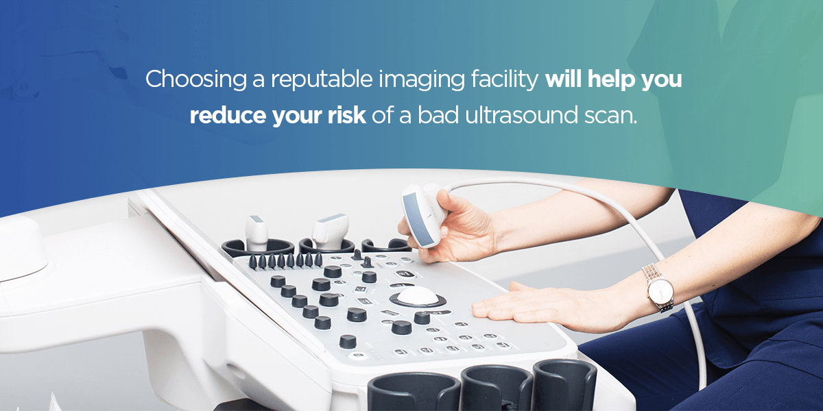
What are the limitations of ultrasound imaging?
Because sound waves can’t pass through certain objects, ultrasound isn’t an effective imaging technique for anything blocked by bone, gas or fat, such as the inside of your stomach or lungs. You’ll typically need a different type of exam to get a clear picture of these structures. Additionally, failure to follow the recommended pre-scan protocol can make it more difficult for the technologist to get a clear picture. Some of the most common signs of a bad ultrasound image include:- Incomplete image: An incomplete picture can cause your provider to miss crucial details in the examination area.
- Lack of clarity: Your ultrasound image should be clear, with distinct shapes and boundaries between different structures. An unclear image is significantly harder to interpret and diagnose.
- Artifacts: The presence of artifacts — unwanted elements like shadows or lines — in an ultrasound scan can make your results harder to interpret.
Why are ultrasound pictures so important?
Diagnostic ultrasound scans give physicians a way to examine internal structures in real time, which can significantly reduce the time needed to diagnose serious conditions such as:- Infections
- Uterine polyps and fibroids
- Tumors and cysts
- Blocked blood vessels
- Thyroid conditions
- Cardiovascular issues
- Monitor the baby’s growth
- Check the baby’s position
- Discover the baby’s sex
- See if there is more than one baby
- Screen for birth defects or genetic disorders
- Check amniotic fluid levels and placenta position
- Guide test sample collection
- Determine the approximate due date
How does an ultrasound work?
Unlike x-rays or magnetic resonance imaging (MRIs), ultrasound scans don’t need radiation or magnetic fields to produce images. That’s one of the biggest reasons why they are so safe. Instead, a sonogram uses high-frequency sound waves to quickly create a picture of the structure under examination. Here’s what a typical ultrasound exam looks like:- First, you’ll lie down on the exam table. You may need to change into a hospital gown to give the technologist better access to the area.
- If you’re getting an external scan, the technologist will apply a thin layer of cold gel to the area. This gel is important because it replaces the air surrounding the area, which provides a path for the ultrasound waves to travel into the body faster.
- The technologist will begin the scan by passing the transducer, a handheld probe that resembles a wand, over or into the area. Although you won’t be able to hear them, the transducer will emit high-frequency sound waves into your body.
- The sound waves will rebound off your internal structures and return to the transducer, which converts them into electrical signals.
- The computer translates these signals into images or videos, which it displays on a nearby monitor. You’ll be able to see them from the exam table if you’re undergoing a pregnancy ultrasound.
Are there any risks to ultrasounds?
Ultrasound scans are generally safe, but there are a few things you should be aware of before you schedule an appointment:- Allergic reaction: Many transducers have a latex-covered head, which can make internal scans dangerous for people with latex allergies. If you have a latex allergy, notify your provider ahead of time so they know to use a different probe cover.
- Heat: While prolonged exposure to ultrasonic waves can cause your body tissues to heat, this temperature change poses no risk to you.
- Cavitation: In some cases, an ultrasound exam can create tiny gas bubbles in your body tissues. While cavitation poses no immediate risk to you or your baby, experts are still unsure whether there is a long-term effect.
- Keepsakes: Some non-medical businesses offer pregnant mothers the opportunity to take a 3D or 4D ultrasound “keepsake.” Medical professionals have warned against these scans, as inexperienced operators can expose the baby to unnecessary risk.
How to get the best ultrasound pictures
Preparation protocols will look different depending on the type of ultrasound you need. These precautions help ensure your technologist can get a clear view of the area being examined. For a scan of the pelvic region or urinary tract, your provider will typically ask you to come in with a full bladder. They will typically recommend you drink between 18 to 32 ounces of water 30 minutes to an hour before your appointment. This extra fluid allows the sound waves to travel faster through your body, which helps create a clearer, more detailed image. Preparations for pregnancy ultrasounds are usually similar to those of pelvic scans. Eat normally and drink plenty of water before your appointment. Although you’ll likely need to change into a gown, arriving in comfortable clothing may help you feel more at ease while you wait. For transvaginal, abdominal or inferior vena cava (IVC) ultrasounds, you’ll likely need to come in fasted with an empty bladder. A full stomach or bladder can obstruct the technologist’s view of the other structures they need to examine, which can negatively impact results. Relaxation tips for good ultrasound pictures
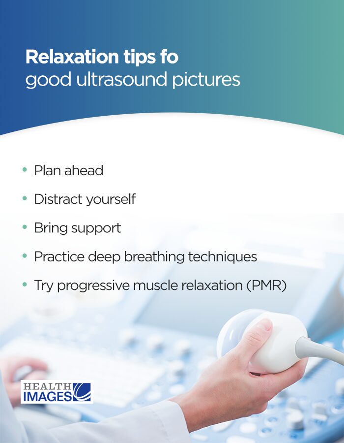
Fidgeting or holding tension in your muscles can make it harder to get clear ultrasound pictures. If medical settings tend to make you anxious, finding ways to relieve your stress can help ensure the best ultrasound results:
 Relaxation tips for good ultrasound pictures
- Plan ahead: Making preparations before your appointment can help reduce your stress and anxiety by ensuring you arrive on time with everything you need.
- Distract yourself: Bringing along books or games can help you keep your mind busy in the waiting room so you can keep the anxiety at bay.
- Bring support: Ask a reliable friend or family member to come with you for emotional support. While they may not be able to be in the exam room with you, they can help encourage you before the test.
- Practice deep breathing techniques: Consciously calming your breathing can help you clear your mind and relax your body. The 4-7-8 technique is an effective method that involves four seconds of breathing in, seven seconds of holding your breath and eight seconds of breathing out.
- Try progressive muscle relaxation (PMR): This relaxation technique involves tensing your muscles and slowly releasing them in a progression from head to toe. While many people use it to help them get to sleep, it’s also effective for relieving symptoms of general anxiety.
- Talk through fears: Explain to your child what will happen during their visit, and let them talk to you about what’s worrying them. If they’re having trouble explaining what they’re afraid of, try asking questions to help them out.
- Validate their feelings: As a parent, your natural temptation is to tell your child that there’s nothing to worry about — but it’s also important to avoid shutting expression down. Let your child know you understand their feelings and reassure them you’ll be right there during the test.
- Bring distractions: Bringing along a favorite toy or book can help keep your child’s mind occupied before and during the exam, which will help relieve some anxiety.
3D vs. 4D ultrasound: What’s the difference?
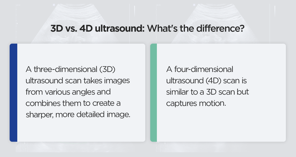 Traditional ultrasound images are two-dimensional (2D), which means they produce flat images of the structure being examined. Although these images are the medical standard for most conditions, including prenatal scans, there are instances in which you may need a better view.
A three-dimensional (3D) ultrasound scan takes images from various angles and combines them to create a sharper, more detailed image, almost like a photograph. In pregnancy ultrasounds, a 3D scan lets you see some of your baby’s facial features and, sometimes, their fingers and toes. You’ll usually receive this type of scan around 24 to 34 weeks if your provider offers it.
A four-dimensional ultrasound (4D) scan is similar to a 3D scan but captures motion. In a 4D prenatal scan, for example, you can see the baby kicking, sucking their thumb or opening their eyes in real time.
That said, 3D and 4D ultrasound technology is still fairly new. Although many clinics are adopting this technology, there are still only a few medical circumstances that would require you to get a 3D or 4D scan:
Traditional ultrasound images are two-dimensional (2D), which means they produce flat images of the structure being examined. Although these images are the medical standard for most conditions, including prenatal scans, there are instances in which you may need a better view.
A three-dimensional (3D) ultrasound scan takes images from various angles and combines them to create a sharper, more detailed image, almost like a photograph. In pregnancy ultrasounds, a 3D scan lets you see some of your baby’s facial features and, sometimes, their fingers and toes. You’ll usually receive this type of scan around 24 to 34 weeks if your provider offers it.
A four-dimensional ultrasound (4D) scan is similar to a 3D scan but captures motion. In a 4D prenatal scan, for example, you can see the baby kicking, sucking their thumb or opening their eyes in real time.
That said, 3D and 4D ultrasound technology is still fairly new. Although many clinics are adopting this technology, there are still only a few medical circumstances that would require you to get a 3D or 4D scan:
- Getting a better look at facial and skeletal abnormalities, such as cleft lip
- Monitoring a high-risk pregnancy
- Evaluating complex gynecological conditions like uterine polyps anzd fibroids
Schedule your ultrasound with Health Images today
Do you need an ultrasound scan? Consider scheduling your appointment at one of our state-of-the-art outpatient imaging centers. Our expert technologists provide fast, accurate results without sacrificing patient comfort and convenience. So whether you’re nervous about coming in for your first ultrasound during pregnancy or you want to accompany your child during a scan, our staff will provide the safe, compassionate care you need. Discover what makes us the best choice for outpatient imaging procedures. Visit our locations page to find your nearest imaging center and schedule your appointment. Questions? Please read our frequently asked questions (FAQs) page to learn more about what to expect.